X-Ray, MRI, or CT Scan?
How is Diagnostic Imaging Used for Back Pain?
You trust SpineOne to diagnose and treat your back, neck, and joint pain. So how does that process begin, and what role, if any, does diagnostic imaging play in the process? The doctors at SpineOne will conduct an examination of your range of motion, nerve function and the area of discomfort. There are many ways our doctors will determine the cause of your pain
If your doctor needs to examine your body’s internal structure to get a deeper understanding of your condition, or pinpoint the injured or inflamed structure at the root of your condition, they may recommend you for one of three types of diagnostic imaging: magnetic resonance imaging (MRI), computed tomography (CT) scans, or X-rays. These are different means of examining your body’s internal structure to get a deeper understanding of the cause of your pain or illness. In this article we’ll discuss the three imaging modalities, and their role in diagnosing back pain.
Do I Need an MRI for My Back Pain?
For the most common types of acute back pain – that is pain that lasts from a few days to a few weeks – diagnostic imaging may not be required. This type of pain can often be diagnosed with a physical examination and may be treated using conservative methods such as rehab and medication. Our doctors will typically only order imaging for persistent symptoms, or certain “red flags” that are revealed during an examination. The red flags include:
- Severe or progressive neurological issues
- Sudden back pain with spinal tenderness
- A serious underlying condition
- Fever
- Trauma

The Role of Medical Imaging in Diagnosing Back Pain
Diagnostic imaging, including MRI, CT, and X-Ray, is used to diagnose a range of spinal conditions from mild to severe. Spinal abnormalities, such as herniated discs, are frequently diagnosed using medical imaging. Other back problems that can be diagnosed using medical imaging include:
What is an MRI?
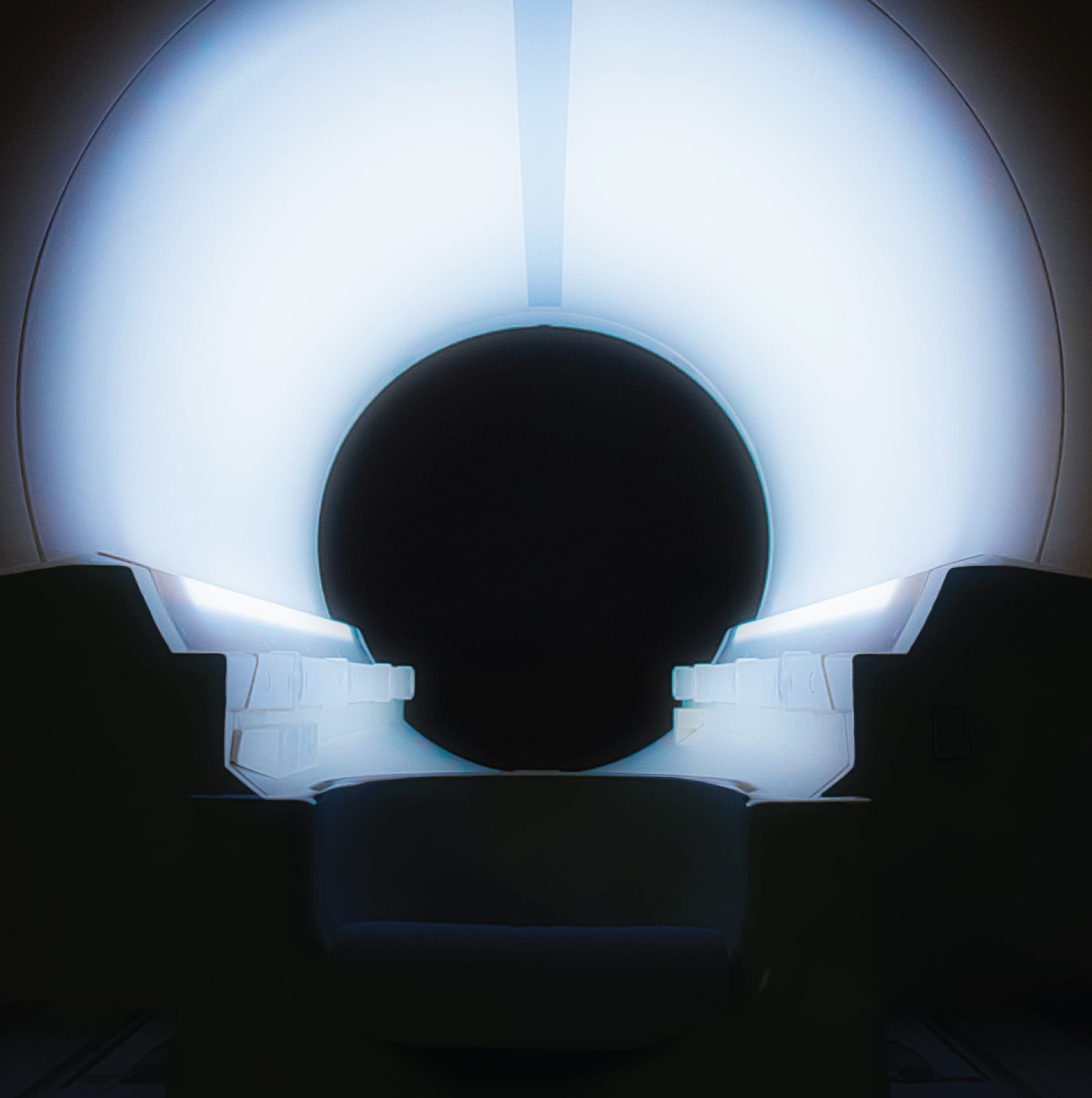
MRIs (Magnetic Resonance Imagers) create a magnetic field in your body and sends radio waves to the area being examined. The process causes fat and water molecule protons in your bone and tissue to resonate, showing a detailed computer image. This results in a detailed, cross-sectional image of the body’s internal structure. With this image, your doctor can get a clear visual of your bones, joints, ligaments, blood vessels, cartilage and discs.
What is a CT Scan?
A CT scan (Computed Tomography) creates a three-dimensional, cross-sectional image using specialized X-ray technology. This scan is often pronounced as “cat scan,” and created a 360-degree visual showing detailed structures in your body. The test is often broken down into “slices” – each slice, or section, is scanned separately for a variety of health conditions.
What is an X-Ray?
X-ray testing is more commonly used than the other medical imaging technologies. This modality is commonly used in dentist offices and chiropractors. An X-Ray machine sends low levels of electromagnetic radiation through the body to create a 2D image of your skeleton. When the rays hit a dense object, like bone, it appears as a white structure on the image. These images are used to check for structural issues like osteoporosis, degeneration, fractures and alignment problems.
Why Have All Three at SpineOne?
SpineOne’s clinic, where patient exams are performed, includes an X-Ray where most of our patients get a quick scan to help the doctor get a look at any skeletal issues that may be contributing to your condition.
Two floors down from the SpineOne clinic is our imaging center, Park Meadows Imaging. This center houses modern MRI and CT scanners, and is where our doctor will send you if he needs a more detailed look at the nerves, muscles, and soft tissue in a specific area.
The SpineOne medical center is one of few spine specialists in the area to house all three imaging modalities in one location. Why do we do it? Because sending patients to a specialist imaging center for scans would interfere with our mission to provide urgent care for back, neck, and joint pain. The cycle that exists at most facilities – referral, waiting for an appointment, having the images read, and finally developing a treatment plan – can leave a patient struggling with pain in their day-to-day lives for weeks, sometimes months.
By having imaging services in the same building as our exam and treatment centers we can reduce, sometimes remove, the need for multiple trips. Our specialists are available to answer your questions about our imaging services or same-day appointment options.


